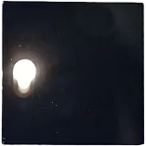
Multimodal Super‐Resolution Imaging of Nitrogen‐Vacancy Centers via High‐Index‐Induced Structured Illumination Microscopy and Optically Detected Magnetic Resonance Spectrometry (Adv. Sci. 24/2026)
Paper details
Section titled “Paper details”| Field | Value |
|---|---|
| Journal | Advanced Science |
| Published | 2026-04-01 |
| Authors | Kyu Ri Choi, Mohammed Zia Jalaludeen, Samuel Begumya, Yan Qiu Du, Dong Hee Park |
| DOI | 10.1002/advs.75192 |
Speckle-assisted structured illumination microscopy resolved two NV-center features separated by approximately 98 nm. The emitters were about 100 µm beneath the surface of type 1b HPHT diamond. The authors report more than twofold improvement over diffraction-limited imaging and simultaneous ODMR spectra with local magnetic-field estimates.
Resolution compared with diffraction-limited imaging
Section titled “Resolution compared with diffraction-limited imaging”Diffraction-limited imaging merged NV ensembles separated by less than approximately 360 nm. Blind structured illumination microscopy reconstruction separated emission peaks that appeared merged in the wide-field image.
| Region | Reported feature separation | Blind-SIM result |
|---|---|---|
| c | approximately 300 nm | Two peaks clearly resolved |
| d | approximately 170 nm | Features resolved |
| e | approximately 150 nm | Features resolved |
| f | approximately 98 nm | Two features distinctly resolved |
The 98 nm pair was the strongest reported result. The authors describe it as more than twofold improvement compared with the diffraction limit. Conventional linear SIM typically yields a twofold resolution improvement, according to the paper’s cited literature. The reported gain therefore exceeds the twofold gain cited for linear SIM.
High-index speckle formation
Section titled “High-index speckle formation”Random speckle patterns entered diamond from air, where n = 1. Diamond has a refractive index of approximately 2.4. This index compressed low-spatial-frequency illumination into finer real-space features and broader transverse spatial-frequency content.
FDTD simulations compared electric fields in both media. Their Fourier spectra were bounded by k0 = 2π/λ in air and extended to nk0 in diamond. The simulated field therefore reached components beyond the free-space cutoff. These components modulated NV emission at sub-diffraction scales and supplied information for reconstruction.
Prior high-index SIM approaches used metamaterials, plasmonic structures, polymer films, epsilon-near-zero membranes, or photonic chips. The authors set diamond apart because it is already the host required for NV imaging.
NV-center preparation in bulk diamond
Section titled “NV-center preparation in bulk diamond”The centers were created in type 1b HPHT diamond by 780 nm femtosecond laser irradiation. A volumetric Z-stack placed the enhanced-emission interface about 100 µm beneath the surface. The authors attribute the emission increase to GR1 neutral vacancies recombining with nearby substitutional nitrogen. This produced NV centers within the focal volume.
The laser writing occurred under ambient conditions without high-temperature annealing. The authors state that it reduces collateral damage and graphitization.
Arbitrary patterns could be inscribed in the interface layer. The resolution test used pairs of 200 nm × 200 nm square irradiation regions. Their designed center-to-center spacings were 250 nm, 300 nm, and 400 nm. NV formation was stochastic, so individual emitters inside each square could lie closer than the design spacing. That variability allowed reconstructed pairs to have separations below the nominal spacing.
Multimodal acquisition and reconstruction
Section titled “Multimodal acquisition and reconstruction”The negative NV center has a triplet ground state with sublevels ms = 0 and ms = ±1. Its zero-field splitting is Dgs = 2.87 GHz. The ms = 0 state emits more strongly under optical excitation. The ms = ±1 states have an approximately 20% lower photon emission rate. This contrast allows RF-driven spin transitions to modulate fluorescence.
A 532 nm CW laser had approximately 160 mW output. An AOM selected the first-order beam before coupling it into a multimode fiber. Mechanical fiber deformation and a motorized fan generated changing speckle patterns. A 60×, 0.9 NA objective projected them through a 512 to 536 nm excitation filter.
The imaging objective also collected NV fluorescence through a 600 nm long-pass filter. A loop antenna applied RF, while a magnet supplied the static field. An sCMOS camera recorded synchronized images.
For each static speckle pattern, the system acquired a full RF sweep under RF-on and RF-off conditions. Blind-SIM jointly estimated the effective illumination and sample distribution from a selected frame subset. ODMR modulation added spin-dependent contrast to the spatial speckle data. The authors describe this as dual modulation of the reconstruction.
Spatially resolved magnetic-field mapping
Section titled “Spatially resolved magnetic-field mapping”To retrieve local fields, the authors divided the wide-field image into grid tiles. Pixel intensities across the image stack formed an ODMR spectrum for each tile. The outermost resonances had the largest Zeeman splitting and were assumed to come from NV axes aligned with the field.
The field vector came from a Hamiltonian inversion using the upper and lower resonance frequencies. Least-squares fitting and outlier rejection supported the fit. Regions with insufficient signal-to-noise ratio were excluded from the final map.
A four-tile analysis produced six ODMR peaks from three crystallographic orientations. Representative spectra elsewhere showed eight peaks from four orientations. Retrieved vectors were uniform across the wide-field area. Relative field-strength uncertainty stayed below approximately 6%. Angular precision remained within 1 to 3°.
Control reconstructions used speckle alone at an off-resonant RF frequency and the same frame count. The dual-modulation reconstruction separated close emitters more robustly.
Acquisition time and reconstruction burden
Section titled “Acquisition time and reconstruction burden”The full dataset contained 31,320 raw images. It comprised 261 RF frequency points, 20 frames per point, and six repetitions. Each frequency point included ten RF-on and ten RF-off frames. The camera period was 50 ms, corresponding to 20 frames/s, at approximately 60 mW excitation. Full acquisition took approximately 1,566 s, or 26.1 min, excluding minor hardware overhead.
Only 100 to 300 frames from the full stack were required for blind-SIM reconstruction. Once ODMR resonances were known, the authors proposed toggling RF near selected frequencies instead of scanning all 261 points. That reduced protocol would take approximately 5 to 15 s at the current frame period. The reconstruction was performed in MATLAB.
Limits and material selection
Section titled “Limits and material selection”The 98 nm result remains an optical estimate because the emitters lie about 100 µm below the surface. Direct nanoscale ground-truth mapping by SEM or AFM was not accessible. The paper therefore presents the resolution and vector maps as optical- and ODMR-constrained estimates.
The authors did not directly map the compressed speckle field at the diamond interface. Near-field probes or a thin fluorescent probe layer were proposed for future validation. Imaging 100 µm deep with a 0.9 NA air objective also introduced spherical aberration. Blind-SIM and ODMR modulation preserved robust reconstruction in their data. Adaptive optics and immersion objectives were suggested, not tested.
The magnetometry is a proof of concept. Lock-in camera detection was proposed as a route to greater sensitivity and precision. Localization-based methods can offer higher nominal resolution, but they usually require sparse blinking emitters, point excitation, or high-power illumination. They are less suited to dense subsurface NV ensembles in this configuration.
For material selection, the demonstrated platform uses bulk type 1b HPHT diamond and a written NV interface at about 100 µm. A system based on this result therefore needs deep optical access, calibrated RF resonance control, and reconstruction that accounts for spherical aberration.
View Original Abstract
Super-Resolution Imaging In their Research Article (DOI: 10.1002/advs.202522495), Shilong Li, Yeon Ui Lee, and co-workers present a multimodal super-resolution imaging technique for nitrogen-vacancy centers, integrating high-index-induced structured illumination microscopy (SIM) with optically detected magnetic resonance (ODMR) spectrometry. By overcoming traditional diffraction limits, this approach enables high-fidelity spatial mapping and quantum sensing, offering a versatile platform for exploring quantum materials and biological systems with unprecedented precision and sensitivity.

Electronic Grade Intrinsic Diamond Epitaxial Wafer H Terminated Superior 100 Mosfet Ready Substrate
View Specs →
Free Standing Polycrystalline Cvd Diamond Membrane
View Specs →